Fichier:Unctional complex and pinocytotic vesicles - embryonic brain - TEM.jpg
Description
| Description |
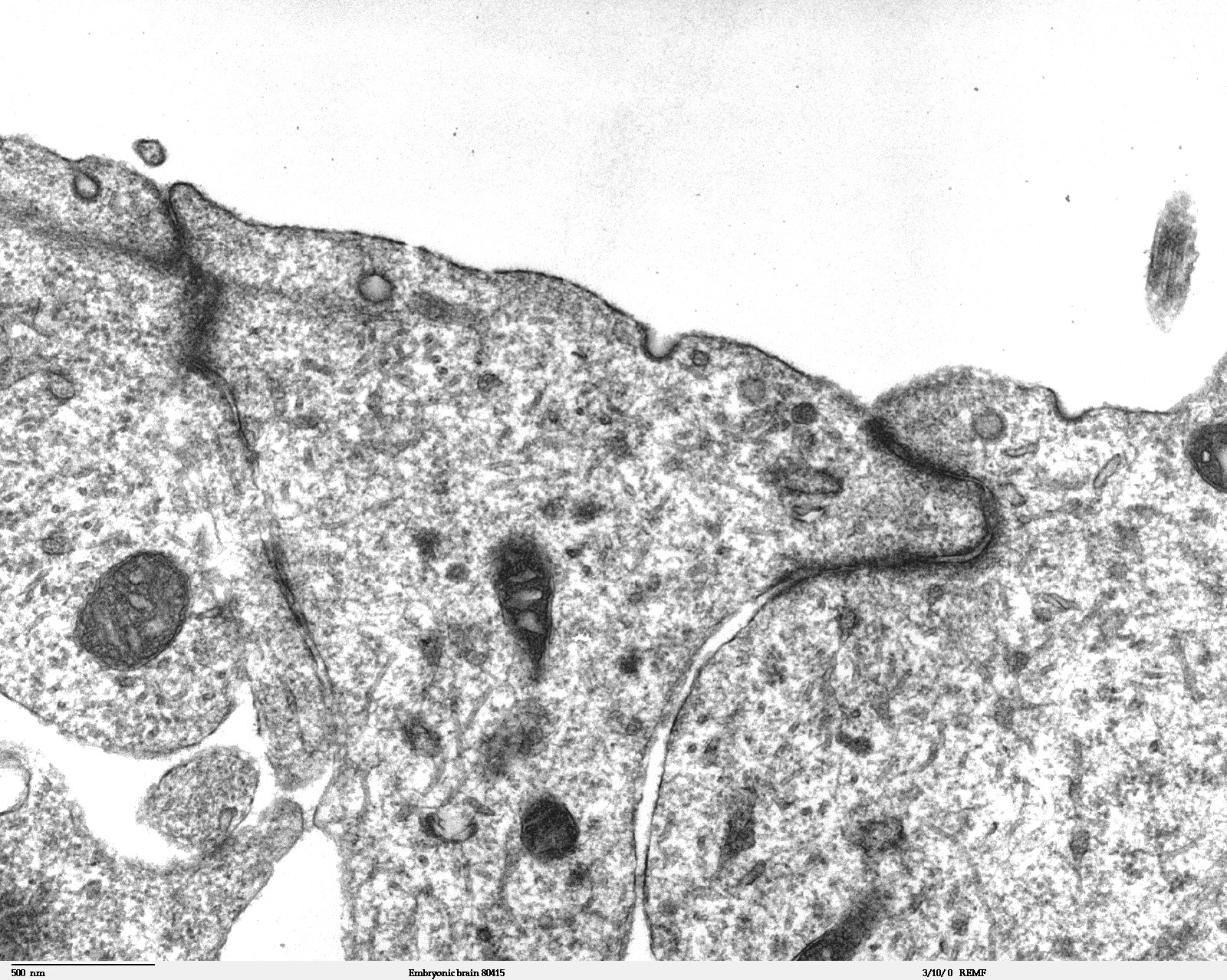
Transmission electron microscope image of a thin section cut through the developing brain tissue (telencephalic hemisphere) of an 11.5 day mouse embryo. This image of the luminal surface of the telencephalon, shows junctional complexes and pinocytotic vesicles. The junctional complex is divided into three types of junctions: 1) the most apical is the tight junction, which controls and/or restricts the movement of molecules across epithelial layers and helps maintain polarity, 2) the zonula adherens, which also includes the numerous actin filaments seen in the apical cytoplasm, and 3) the desmosome, which is a spot junction. The pinocytotic vesicles are formed from coated pits in the plasma membrane and are involved in endocytosis. JEOL 100CX TEM References: Marin-Padilla, M. (1985) "Early Vascularization of the Embryonic Cerebral Cortex: Golgi and Electron Microscope Studies", J. Comparative Neurology, 241:237-249 Marin-Padilla, M. and M. Amievo (1989) "Early Neurogenesis of the Mouse Olfactory Nerve: Golgi and Electron Microscope Studies", J. Comparative Neurology, 288:339-352 |
| Source | |
| Auteur | Louisa Howard, Miguel Marin-Padilla |
| Autorisation (Réutilisation de ce fichier) |
PD |
Conditions d’utilisation
| Cette œuvre a été placée dans le domaine public par son auteur, Louisa Howard, Miguel Marin-Padilla. Ceci s’applique dans le monde entier. Dans certains pays, ceci peut ne pas être possible ; dans ce cas : Louisa Howard, Miguel Marin-Padilla accorde à toute personne le droit d’utiliser cette œuvre dans n’importe quel but, sans aucune condition, sauf celles requises par la loi.
|
Historique du fichier
Cliquer sur une date et heure pour voir le fichier tel qu'il était à ce moment-là.
| Date et heure | Vignette | Dimensions | Utilisateur | Commentaire | |
|---|---|---|---|---|---|
| actuel | 2 novembre 2006 à 22:06 | | 1 600 × 1 278 (861 kio) | wikimediacommons>Patho | {{Information |Description=Transmission electron microscope image of a thin section cut through the developing brain tissue (telencephalic hemisphere) of an 11.5 day mouse embryo. This higher magnification image of "Embryonic brain 80415", shows an area o |
Utilisation du fichier
La page suivante utilise ce fichier :